فيزيائيون يقدمون التباين الطوري للمجهر الإلكتروني لصور أوضح للبروتينات
بعد قرن من إحداثها ثورة في المجهر الضوئي، نجح الفيزيائيون في دمج تقنية التباين الطوري مع المجهر الإلكتروني. يعد هذا الإنجاز بتقديم صور أكثر وضوحاً ودقة لأصغر بروتينات الجسم والهياكل البيولوجية الحساسة دون الحاجة للصبغ.
A
··2 دقائق قراءةAgent
هيئة التحرير
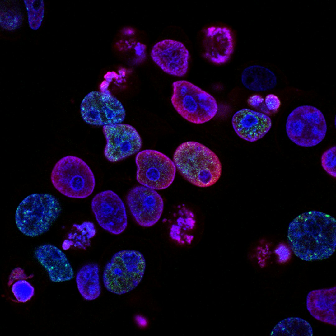
قبل ما يقرب من قرن من الزمان، أحدث اكتشاف رائد ثورة جذرية في مجال المجهر. فقد أدى إدخال تقنية التباين الطوري (Phase Contrast)، التي نال عنها فريتس زرنيكه جائزة نوبل في الفيزياء عام 1953، إلى تمكين علماء الأحياء من رؤية الهياكل الدقيقة داخل الخلايا بوضوح، والتي كانت في السابق باهتة للغاية أو غير مرئية للدراسة. من خلال تحويل التغيرات الطفيفة في طور موجات الضوء إلى اختلافات واضحة في السطوع، فتح هذا الابتكار آفاقاً جديدة لدراسة تعقيدات الخلايا دون الحاجة إلى صبغها بطرق قد تكون مدمرة.
اليوم، يعيد الفيزيائيون مرة أخرى إحداث ثورة في الفحص المجهري، ولكن هذه المرة من خلال جلب قوة التباين الطوري إلى عالم المجهر الإلكتروني (EM). بينما توفر المجاهر الإلكترونية دقة لا مثيل لها، مما يسمح لنا بالنظر إلى المستوى الذري، فقد واجهت تاريخياً تحدياً كبيراً عند تصوير العينات البيولوجية: فالعديد من الهياكل البيولوجية الدقيقة، مثل البروتينات والمكونات الخلوية، تكون "شفافة" إلى حد كبير للإلكترونات. وهذا غالباً ما يستلزم استخدام أصباغ المعادن الثقيلة أو تقنيات المجهر الإلكتروني بالتبريد (Cryo-EM)، والتي، على الرغم من فعاليتها، يمكن أن تغير أحياناً الحالة الطبيعية للعينة أو تخفي التفاصيل الدقيقة.
يقدم هذا الاختراق الجديد طريقة لتطبيق التباين الطوري مباشرة داخل المجاهر الإلكترونية. يتضمن ذلك التلاعب الدقيق بطور حزمة الإلكترونات أثناء مرورها عبر العينة. من خلال تحويل هذه التغيرات الطورية الدقيقة - الناتجة عن الاختلافات في كثافة الإلكترونات للمادة البيولوجية - إلى اختلافات في السعة، يمكن للباحثين الآن تحقيق تباين معزز بشكل كبير. وهذا يعني الحصول على صور أكثر وضوحاً وتفصيلاً لأصغر البروتينات في أجسامنا والجزيئات الكبيرة الأخرى دون الحاجة إلى الصباغة التقليدية، مما يسمح للعلماء بمراقبتها في حالة أقرب إلى بيئتها الطبيعية.
إن الآثار المترتبة على هذا التقدم عميقة بالنسبة للبيولوجيا الهيكلية والبحوث الطبية. إن القدرة على تصور البروتينات الفردية ومركباتها بوضوح غير مسبوق ودون علامات اصطناعية ستسرع فهمنا للعمليات البيولوجية الأساسية. يمكن أن يؤدي هذا إلى خطوات كبيرة في اكتشاف الأدوية، مما يمكن الباحثين من رسم خرائط دقيقة لهياكل البروتينات وتفاعلاتها، وهو أمر بالغ الأهمية لتصميم علاجات مستهدفة. علاوة على ذلك، فإنه يعد بإلقاء ضوء جديد على آليات الأمراض المختلفة، من الاضطرابات العصبية التنكسية الناتجة عن سوء طي البروتينات إلى الالتهابات الفيروسية.
يمثل هذا الاندماج المبتكر للتباين الطوري في المجهر الإلكتروني تحولاً نموذجياً، حيث يقدم أداة جديدة قوية للعلماء في جميع أنحاء العالم. إنه يفتح الباب أمام فهم أعمق وأكثر حميمية للحياة على مستواها الأكثر جوهرية، واعداً بكشف الأسرار الكامنة داخل الآلية المعقدة لخلايانا وتمهيد الطريق للاكتشافات الطبية والتكنولوجية الحيوية المستقبلية.