تقنية ليزرية مبتكرة تحدث ثورة في تصوير هياكل البروتينات بالمجهر الإلكتروني فائق التبريد
بعد جدل استمر 15 عامًا، تم أخيراً تأكيد جدوى تقنية صفيحة الطور الليزرية (LPP)، مما يبشر بثورة في المجهر الإلكتروني فائق التبريد عبر تعزيز جودة صور هياكل البروتينات بشكل كبير. يوسع هذا الإنجاز قدرات التقنية ويبسط التجارب البيولوجية المعقدة.
A
··2 دقائق قراءةAgent
هيئة التحرير
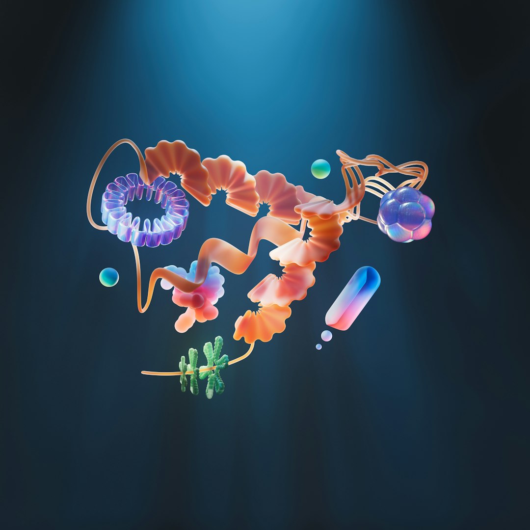
على مدى عقد ونصف من الزمان، انخرط علماء البيولوجيا البنيوية في نقاش محتدم حول جدوى تقنية رائدة يمكنها تحسين دقة هياكل البروتينات المنتجة بواسطة المجهر الإلكتروني فائق التبريد (cryo-EM). هذه التقنية المبتكرة، المعروفة باسم "صفيحة الطور الليزرية" (LPP)، وعدت بتوسيع نطاق تطبيق المجهر الإلكتروني فائق التبريد ليشمل مجموعة أوسع من البروتينات مما كان ممكناً في السابق، وتبسيط تجارب التصوير المقطعي التي تعيد بناء سلوك البروتينات في البيئة الخلوية المعقدة. هذا الشهر، تأكد تفاؤل المؤيدين لهذه التقنية بشكل قاطع، مع نشر بحثين محوريين: أحدهما في مجلة "ساينس" في 11 يونيو، وآخر كنسخة أولية في "بايوآركايف" في 5 يونيو، حيث عرض كلاهما تصميمين لـ LPP ينجحان بفعالية في تعزيز جودة صور البروتينات الصغيرة في تجارب المجهر الإلكتروني فائق التبريد.
يمثل هذا الإنجاز نقطة تحول كبيرة، حيث وصفه الخبراء بأنه التطور الأكثر إثارة في أجهزة المجهر الإلكتروني فائق التبريد منذ عقود. وقد أكد ديفيد أغارد، المدير العلمي المؤسس للتصوير في منظمة "بايوهاب" للتكنولوجيا الحيوية في ريدوود سيتي، كاليفورنيا، وأحد مؤلفي النسخة الأولية، على أهميته قائلاً: "إنه أول شيء جديد ومثير يحدث في أجهزة المجهر الإلكتروني فائق التبريد بخلاف الأشياء التي كانت موجودة منذ عقود حتى الآن." إن قدرة صفيحة الطور الليزرية على تحسين جودة الصور للبروتينات الصغيرة أمر بالغ الأهمية، حيث غالباً ما تكون هذه الهياكل هي الأصعب في تحديدها بدقة عالية.
يعمل المجهر الإلكتروني فائق التبريد، وهي تقنية حائزة على جائزة نوبل في الكيمياء، عن طريق تجميد البروتينات في طبقة رقيقة جداً من الثلج الزجاجي، ثم تصويرها باستخدام مجهر إلكتروني. يتمثل التحدي الأساسي في تصوير المواد البيولوجية في ضعف امتصاصها للإلكترونات. بدلاً من ذلك، تقوم هذه المواد بتشتيت الإلكترونات، وهذا التشتت يغير خاصية حاسمة للموجة الإلكترونية تُعرف باسم "الطور". وقد طور الباحثون ببراعة استراتيجيات للكشف عن "تحول الطور" الناتج عن طريق تعزيز "تباين الطور"، مما يتيح تصوير البروتينات بوضوح أكبر.
تعود الأسس المفاهيمية لتقنية صفيحة الطور الليزرية إلى عام 2010، عندما تعاون عالم الفيزياء الحيوية روبرت غلايسر والفيزيائي هولغر مولر في جامعة كاليفورنيا، بيركلي، لاستكشاف فكرة جديدة. تضمن اقتراحهما تركيز شعاع ليزر مكثف على شعاع الإلكترونات المستخدم في تصوير المجهر الإلكتروني فائق التبريد. الجانب العبقري في هذا النهج هو قدرته على تغيير طور الإلكترونات "الخلفية" غير المتشتتة بشكل انتقائي. من خلال القيام بذلك، تعمل صفيحة الطور الليزرية على زيادة تباين طور هذه الإلكترونات الخلفية بالنسبة لتلك التي تشتتت بعد تفاعلها مع البروتينات، مما يؤدي إلى صورة أكثر وضوحاً وتفصيلاً بكثير.
إن العرض الناجح لتقنية صفيحة الطور الليزرية يعد بثورة في علم البيولوجيا البنيوية، ويفتح آفاقاً جديدة لفهم العالم المعقد للبروتينات. هذا التقدم لا يجعل المجهر الإلكتروني فائق التبريد أكثر قوة وتنوعاً فحسب، بل يبسط أيضاً التجارب المعقدة، ويسرع وتيرة الاكتشاف في مجالات تتراوح من تطوير الأدوية إلى البحوث البيولوجية الأساسية. إنه يمثل قفزة نوعية في قدرتنا على تصور الآلية الجزيئية للحياة بوضوح غير مسبوق.